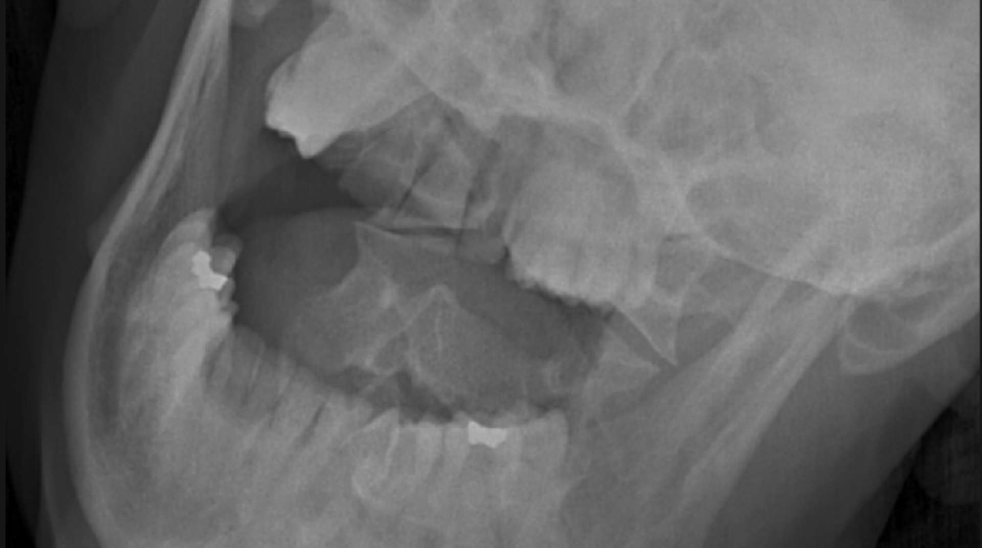
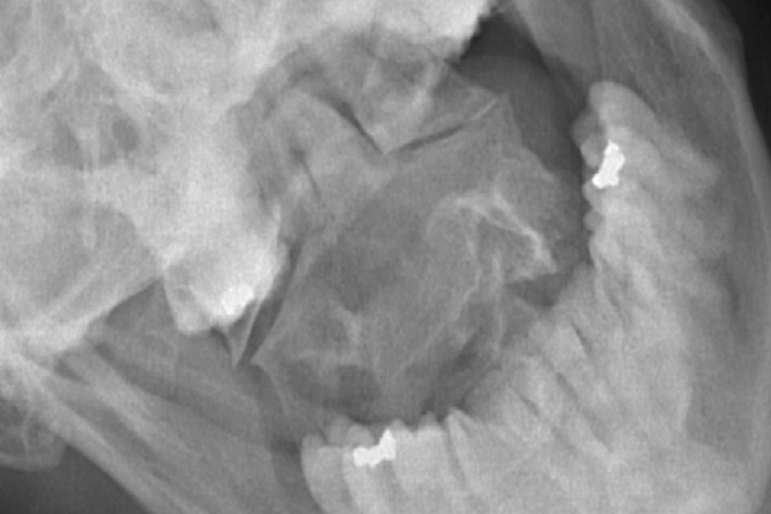
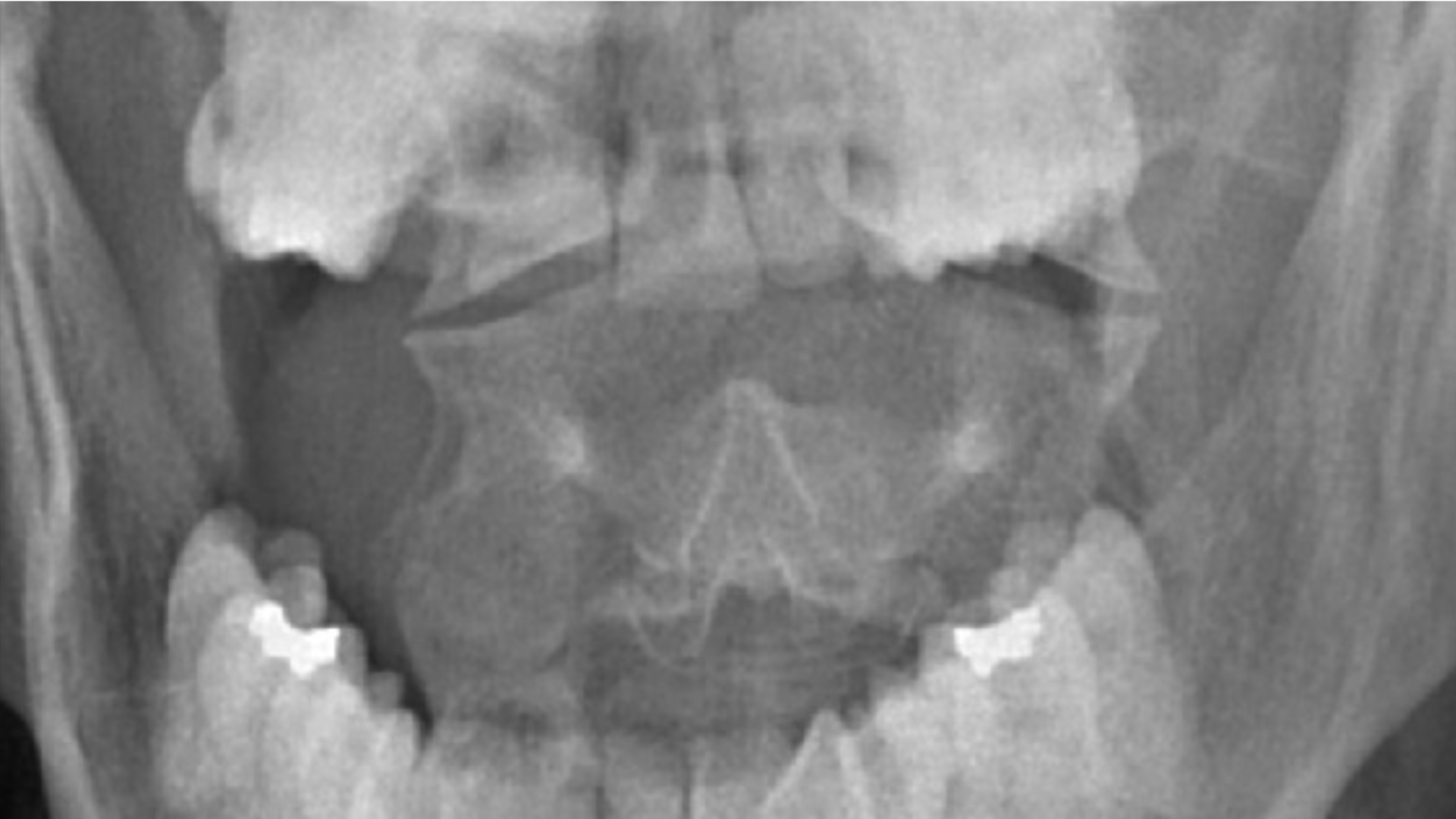
Smartray® GLX 0.1
Smartray® GLX 0.1 is a problem solving patented product with an AI-powered engine developed to uniquely meeting the Gold Standard requirements for Cervicocranial Instability (CCI) diagnosis in the sagittal plane.
By analyzing Pitch, Roll, and Yaw, Smartray® assists healthcare professionals in achieving consistent patient positioning, improving imaging reproducibility, and reducing positioning variability during diagnostic examinations.
The system supports standardized imaging workflows by providing objective positioning guidance before exposure, helping produce consistent diagnostic images across multiple examinations and follow-up studies.
Every examination includes an automated digital positioning report, providing objective documentation that can support clinical review, quality assurance, research, and longitudinal patient follow-up.
Evidence-Based Imaging Evaluation
In September 2024, Smartray® GLX 0.1 imaging data was admitted into evidence in a personal injury case before the Bronx Supreme Court.
The imaging findings contributed to an $800,000 settlement in a case that was about to be lost by the plaintiff.
This represented the first known court admission of Smartray® cervicocranial imaging data as diagnostic medical evidence, highlighting the importance of reproducible and reliable imaging evaluation.
Index #29770/2017e
The Cervicocranium is One of the Most Difficult Anatomies to Evaluate Radiographically
The cervicocranium extends from the skull base through the second cervical intervertebral disk and represents one of the most complex anatomical regions in diagnostic imaging evaluation.
Medical literature also describes the inherent difficulty in obtaining diagnostic frontal and lateral imaging projections because of subtle radiographic findings and complex upper cervical biomechanics.
John H. Harris Jr, MD, DSc.
Radiology 2001; 218:337-351.
Automated Positioning Report
Following each examination, Smartray® GLX 0.1 automatically generates a digital positioning report for every imaging study.
The report includes Pitch, Roll, Yaw, Cephalad Tilt, imaging view, examination time, and study information, providing a permanent record of patient positioning.
Multiple imaging views can be documented within a single examination, allowing clinicians to review positioning across neutral and lateral bending views.
Automated Positioning Report